Aspiration of a Chicken Bone
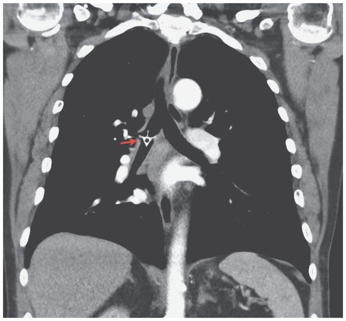
A 78-year-old man presented to the emergency department with stridor, shortness of breath, and fever (38.1°C). Five days earlier, he had come to the emergency department with the sensation of having a foreign body in his throat, approximately an hour after a choking episode that had occurred while he was eating chicken. At that time, the physical examination and plain radiographs of the neck and chest were unremarkable. The foreign body was presumed to have been dislodged, and the patient was discharged home. At the current presentation, computed tomography revealed a high-attenuation structure in the shape of a chicken vertebra in the right mainstem bronchus (arrow). There was minor atelectasis in the right lower lobe without evidence of lobar collapse. Foreign bodies more commonly become lodged in the right side of the bronchial tree than in the left because of its more vertical path and wider lumen. Delayed presentation because of minor symptoms can lead to inflammation and infection. Bronchoscopy was performed, and the chicken bone was successfully removed. The patient recovered well after the procedure and was discharged home on day 3.