


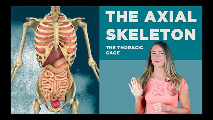
The Thoracic Cage: The Rib cage!
The human thoracic cage consists of the thoracic vertebrae, the ribs, the coastal cartilages and the sternum. They function to protect the contents of the upper abdomen and assist in respiration. A human thoracic cage has 12 ribs on each side that terminate which means form an ending on their anterior or front side into a special cartilage called the coastal cartilage. The upper seven pairs of ribs are called true ribs because they join the medially - which means in the middle and attach directly to the sternum. The remaining five pairs of ribs are called false ribs because their coastal cartilages do not in any way make contact with the sternum directly. Three of the five false ribs link their coastal cartilages with that of the seventh rib and the last two do not attach totally to the sternum. Of the twelve pair of ribs, the last two pairs have no coastal cartilages and appear to be hanging hence called the floating ribs. A normal rib has a shaft which is long and slender having an anterior or frontal and a posterior meaning backside end. It normally goes around the chest and slightly slopes downwards and has an external and an internal surface. The superior which means upper - margin of a rib’s shaft - is very smooth and has a round edge. While the inferior or lower part has a much sharper margin and the internal surfaces’ inferior margin has a coastal groove. The distal end or part situated away from the point of attachment - of the posterior region is enlarged forming a head which provides the articulation site with the facet on an independent vertebra and also simultaneously the head attaches to the body of the superior vertebra higher than the vertebra whose facet is articulated. A facet is a smooth flat circumscribed or circular surface. The rib’s neck is flat and it is laterally positioned, meaning on the side... to the head on which the ligaments attach to. The head of the rib articulates the vertebra’s transverse process with the tubercle. When we say articulation in medical meaning - this is when two bones are attached for the purpose of motion. A rib’s tubercle has a non-articular region that has a rough texture due to ligaments attached to it and an articular region containing a facet which is oval in shape and it corresponds with the facet found on its individual vertebra. Costal cartilages of ribs are made up of a special translucent cartilage called the hyaline cartilage that has expanding and compression capabilities to aid in respiration. The first rib is flat horizontally and has inferior or lower and superior surfaces or upper that are broad. It articulates with the first thoracic vertebrae and has the scalene tubercle on its superior, or upper surface, surface that is distinct. The second rib is two times longer than the first rib and articulates with its thoracic vertebrae same to all the other ribs. The tenth rib only has a single fascet to articulate with individual thoracic vertebrae. A facet is a small flat surface on a bone. Ribs eleven and twelve are distinct and the last ribs and articulate only with their vertebrae’s body and they lack a neck or a tubercle. They are slightly curved, are much shorter than the other ribs and they finish and point anteriorly meaning towards the front. This gives them the characteristic name floating rib since they do not attach to the coastal cartilages.
