


Zunaira saleh11 months ago
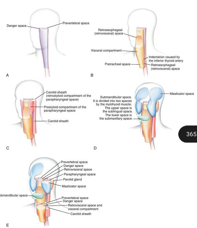
Head and neck in lateral oblique view.
The prevertebral and danger spaces are shown (A), as well as the visceral space (B). Note the separate pretracheal space and retrovisceral space below the level of the insertion of the inferior thyroid artery. The left parapharyngeal space is shown (C), as is the carotid sheath. (D) Masticator and submandibular spaces. (E) Axial cuts through the levels of the upper pharynx and mid-neck illustrating their special relationships. (From Som PM, Curtin HD. Head and Neck Imaging. Philadelphia: Elsevier; 2011:2203–2234.)
Other commentsSign in to post comments. You don't have an account? Sign up now!
