


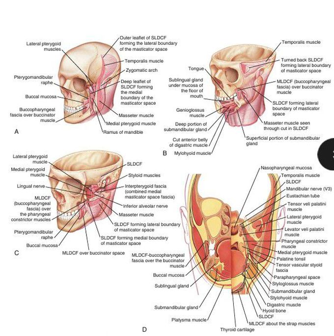
Left oblique view of facial region and neck.
(A) A coronal cut has been made through the ramus of the mandible. The deep and superficial fascial leaflets of the masticator space are shown, as is the relationship of the masticator space to the buccopharyngeal fascia. (B) An axial cut has been made through the temporalis muscle, and a portion of the mandible has been removed to expose the floor of mouth structures. (C) An axial cut has been made through the mid-mandibular ramus. The left zygoma and most of the arch have been removed to expose the deep structures of the region. (D) A coronal drawing of the facial region and upper neck. On the right side of the drawing, the coronal cut is through the region of the foramen ovale and palatine tonsil. On the left side of the drawing, the cut is through the region of mid floor of mouth. MLDCF, Middle layer, deep cervical fascia; SLDCF, superficial layer, deep cervical fascia. (From Som PM, Curtin HD. Head and Neck Imaging. Philadelphia: Elsevier; 2011:2203– 2234.)
