


Zunaira saleh12 months ago
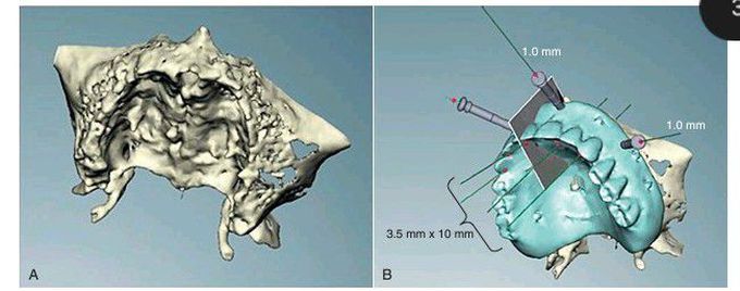
Computer-assisted virtual treatment planning.
(A) Three-dimensional view of the maxilla created from cone-beam computed tomography data. (B) “Virtual” prosthesis placed over the maxillary anatomy. The ideal position and angulation of implant placement can be determined. Individual crosssections can be evaluated.
Other commentsSign in to post comments. You don't have an account? Sign up now!
Related posts
Root canal treatmentComputerized imaging for dentofacial surgical treatment planningCystic Fibrosis TreatmentCystic Fibrosis TreatmentImplant Depth- Anterior TeethPatent Ductus Arteriosus- TreatmentEpidermolysis Bullosa-TreatmentAcromegaly - TreatmentCholelithiasis TreatmentErythema Multiforme Treatment
