


Paget's Or Metastatic Tumor Of The Prostate - Everything You Need To Know - Dr. Nabil Ebraheim
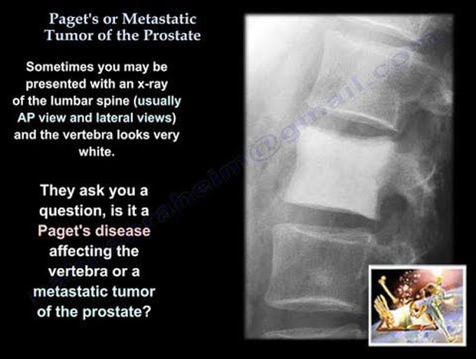
Sometimes you may be presented with an x-ray of the lumbar spine (usually AP view and lateral views) and the vertebra looks very white. The patients will require other tests to differentiate between Paget’s and metastatic tumor. In Paget’s disease, the vertebra looks like a picture frame, and it is called “picture frame vertebra body”. “Picture frame vertebra body” is a radiologic appearance, where the cortex or the vertebral body is thickened. There will be vertebral body expansion and coarsened trabeculae. There is increased opacity of the cortex on all sides of the vertebral body, which is in contrast to the Rugger-Jersey spine, where the sclerosis is seen only at the superior/inferior vertebral endplate. There will be disorganized, new cortical bone formation after excessive osteoclastic activity that causes resorption of the normal bone. You will see condensation of bone along its peripheral margins that resembles a picture frame. Metastatic prostate has an osteoblastic lesion. A metastatic breast tumor can also have an osteoblastic lesion. Paget’s disease can also appear to have an osteoblastic lesion. In Paget’s, there will be an increase in the alkaline phosphatase, an increase in the urinary hydroxyproline and in the N-telopeptide. In general, Paget’s will have a mosaic pattern and also cement lines under the microscope. You can also see the cortical thickening in Paget’s disease. If you see on an x-ray an osteoblastic lesion of the lumbar vertebra in an older, male patient or if you see it on a CT scan, then you are probably dealing with a metastatic prostate carcinoma. When you see the white vertebra in the examination or in the clinic, sometimes this is called the “ivory vertebra sign”. There will be diffuse and hematogenous increase in the opacity of the vertebral body. The vertebral body is normal in size and contour with no change in the adjacent intervertebral discs (they are normal). In adult males, this indicates metastatic cancer of the prostate or it can be breast cancer in females. The histology will show adenocarcinoma with gland formation. The histology and x-rays will establish the diagnosis and the Prostate-Specific Antigen (PSA) will confirm the diagnosis. Paget’s may be polyostotic and may be similar to prostatic metastatic carcinoma and the x-ray will probably show coarsening of the trabeculae pattern rather than an osteoblastic infiltrating process. There is another entity caller Rugger-Jersey spine. This occurs due to hyperparathyroidism. Rugger-Jersey spine describes the prominent sub-endplate density at multiple continuous vertebra levels. There will be an alternating sclerotic, lucent appearance which looks like the horizontal stripes of a rugby jersey. Because Rugger-Jersey spine occurs with hyperparathyroidism, there will be an increased resorption due to excessive parathyroid hormone secretion with subsequent loss of bone mass (why you see the lucent areas). The osteoblasts make osteoid that does not have hydroxyapatite and does not appear dense on x-rays. The areas which are dense or sclerotic at the superior and inferior endplate of the vertebral body indicates accumulation of excess osteoid, and it appears dense when compared to the normal bone.
