


Medvisionover 6 years ago
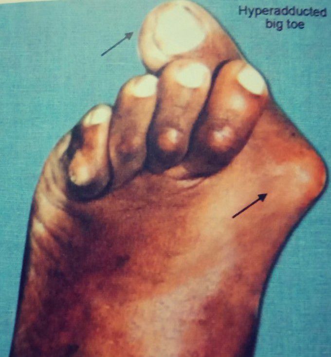
Hallux valgus - most common forefoot deformity
Hallux valgus refers to the deformity in which the big toe passes transversely behind the second toe and there is abnormal prominence of head of first metatarsal bone on the medial side of the foot just behind the big toe. The exposed and prominent head of first metatarsal tends to rub on the shoe ,giving raise to an adventitious Bursa called bunion. The patient experience pain and fatigue during routine 🚶 walking. Causes are arthritis, wearing tight shoes.
over 6 years ago
In initial phase brace can be used to limit the movement of joint. For complete correction surgery should be done.
Other commentsSign in to post comments. You don't have an account? Sign up now!
over 6 years ago
In the beginning you can try physiotherapy and corrective pads for shoes.
