


AshleyCooperabout 8 years ago
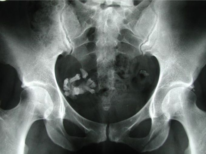
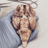
Ovarian Teratoma with Teeth
Ovarian teratomas include mature cystic teratomas (dermoid cysts), immature teratomas, and monodermal teratomas (eg, struma ovarii, carcinoid tumors, neural tumors). Most mature cystic teratomas can be diagnosed at ultrasonography (US) but may have a variety of appearances, characterized by echogenic sebaceous material and calcification. At computed tomography (CT), fat attenuation within a cyst is diagnostic. At magnetic resonance (MR) imaging, the sebaceous component is specifically identified with fat-saturation techniques.
Other commentsSign in to post comments. You don't have an account? Sign up now!
