Feed
Library
Learning
Discuss
Leaderboard
Journal
Sign in
Login
Sign up
Sign in
Feed
Library
Learning
Discuss
Leaderboard
Journal
Yash Kumar
over 6 years ago
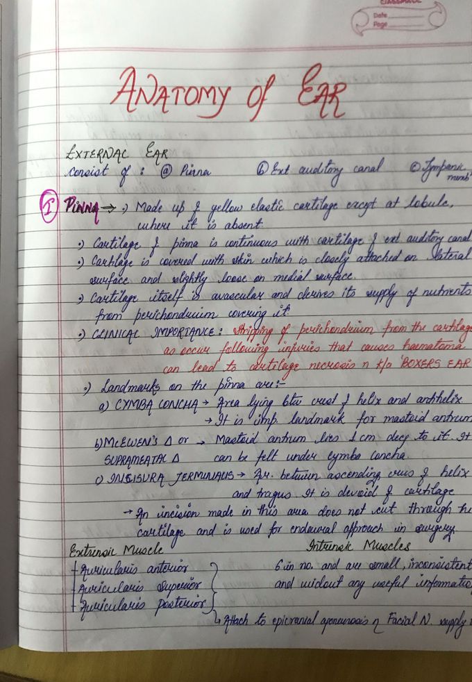
Anatomy Of EAR
It tells about the ear pinna ...part of external ear
Anatomy
ENT
Ear
Yashkumar
1
0
2
Other comments
Sign in
to post comments. You don't have an account?
Sign up now!
Related posts
Left oblique view of facial region and neck.
Head and neck in lateral oblique view.
DNA vs RNA - Differences in Form and Function | Stated Clearly
SternocleidoMastoid Muscle
Neck Anatomy - Organisation of the Neck - Part 1
Sublingual gland anatomy
Brain Blood Supply
Anatomy of Oral Cavity
Neck Muscles
Recent MCQs
Show more MCQs
Recent flashcard sets
Show more flashcards
Feed
Learn
Add
Search
Discuss