


Infected Urachal Cyst
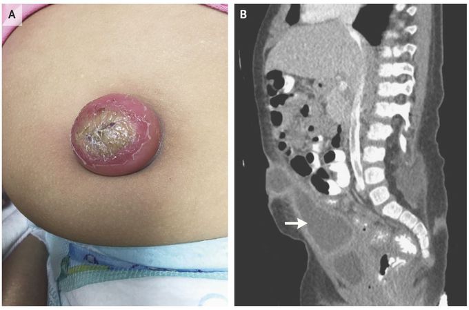
A 19-month-old girl with a history of myelomeningocele and Chiari malformation type II presented with abdominal pain. The initial abdominal examination was normal; however, within 24 hours, the umbilicus became protuberant and erythematous (Panel A). The white-cell count was elevated, with a large proportion of neutrophils. Computed tomography of the abdomen revealed a cystic mass, measuring 6.1 cm by 2.0 cm, which extended from the umbilicus to the dome of the bladder (Panel B, arrow). The mass had no connections with the bladder or umbilicus; therefore, it was diagnosed as an urachal cyst. Antibiotic agents were administered, and purulent material was drained from the infected cyst. Escherichia coli and coagulase-negative staphylococcus grew. Antibiotic treatment was tailored to these organisms. After drainage and a course of antibiotics, the urachal cyst was excised. The differential diagnosis for this condition includes patent urachus and a partial urachal remnant. A patent urachus connects the bladder and umbilicus, resulting in urinary drainage through the umbilicus; a partial urachal remnant connects the urachal sinus to the umbilicus or the urachal diverticulum to the bladder. More than 1 year after surgical correction, the patient had an apparently unrelated infraumbilical abscess requiring incision and drainage, but she has been otherwise well. Nicholas Potisek, M.D. James Weihe, M.D. Wake Forest School of Medicine, Winston-Salem, NC source: nejm.org
