


Bitot’s Spots
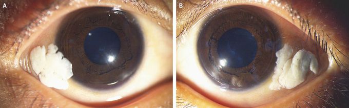
A 4-year-old boy was brought by his father to the ophthalmology clinic with a 1-year history of enlarging white deposits in both eyes and decreased night vision. On examination, the conjunctivae of both the right eye (Panel A) and the left eye (Panel B) appeared dry and wrinkled, with foamy, cream-colored deposits near the outer corners. The corneas were clear, the fundi were normal, and the visual acuity was 20/30 in both eyes. The child appeared pale, with hypopigmented hair, a weight of 10.5 kg (z score of less than −3), and a height of 92 cm (z score of −2.8). The ocular findings were consistent with Bitot’s spots, which are accumulations of keratin, often intermixed with an overgrowth of Corynebacterium xerosis, that result from epithelial metaplasia caused by vitamin A deficiency. Vitamin A deficiency can also cause blindness, as a result of corneal ulceration with scarring, and particularly night blindness, as a result of dysfunction of rod photoreceptor cells. In this patient, the serum vitamin A level was 16.8 μg per deciliter (0.59 μmol per liter) (reference range, 20 to 40 μg per deciliter [0.70 to 1.40 μmol per liter]), and the retinol-binding protein level was 0.01 g per liter (reference range, 0.03 to 0.06). Further history taking revealed extreme poverty, and further examination showed signs of dietary deficiency, with no evidence of intestinal parasites. The patient received an oral dose of vitamin A, which was repeated 4 weeks later. Artificial tears were also administered. The Bitot’s spots abated, although they did not completely resolve, over the course of 12 weeks. The parents were advised to administer another dose of vitamin A every 6 months until the child was 5 years of age. Jagat Ram, M.S. Jitender Jinagal, M.S. Post Graduate Institute of Medical Education and Research, Chandigarh, India source: nejm.org
