
Zunaira salehabout 1 year ago

Hydatid cyst
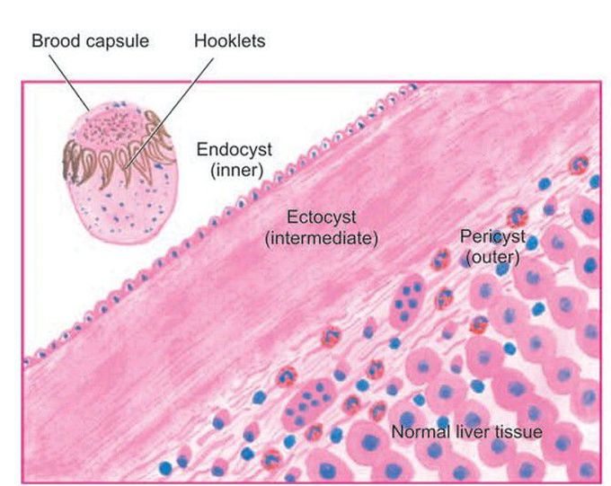
Microscopy shows three layers in the wall of hydatid cyst. Inbox in the right photomicrograph shows a scolex with a row of hooklets.
Other commentsSign in to post comments. You don't have an account? Sign up now!

