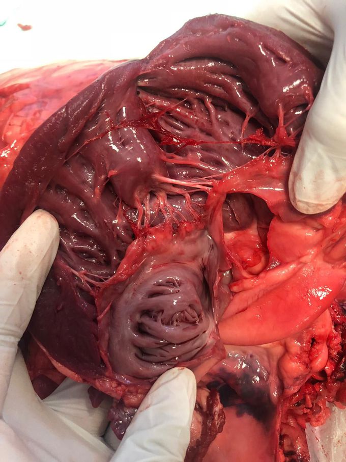
From what I see, I could distinguish the papillary muscles (where your left forefinger is), the threads that form the tricuspid valve; the chordae tendineae, the pectinate muscles (above your left thumb). As a whole picture you are showing the right ventricle of the heart and a little bit of the right atrium (which would consist of the pectinate muscles and orifice of the inferior vena cava), I might be missing the orifice of the inferior vena cava (I believe it's near to your right thumb). I might miss some structures.
I can see the right ventricle, a bit of the right atrium, papillary muscles and some pillars (or we call it that way in France)
It's a papillary muscles, Chordae tendineae which are fibrous cords r attached to come shaped projections of ventricular wall, papillary muscles. The chorale standings prevent the bicuspid n tricuspid valves collapsing bck into the atria during powerful ventricular contraction. Bt according to images the papillary muscles are being shown in right ventricle.


