


Anatomy of Oesophagus
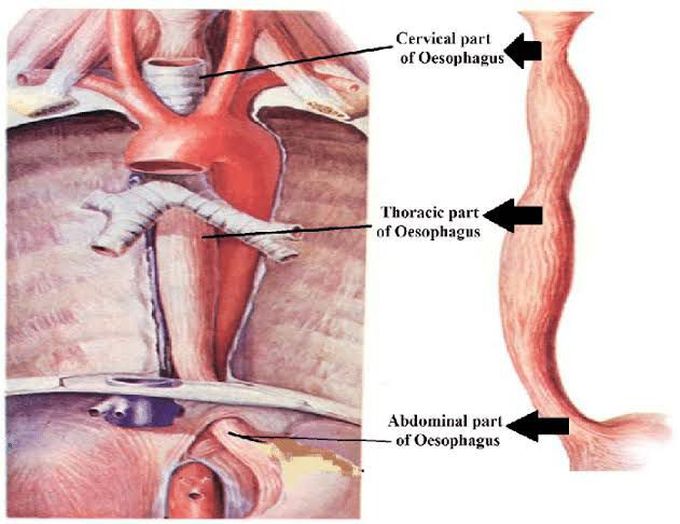
The esophagus (oesophagus) is a 25 cm long fibromuscular tube extending from the pharynx (C6 level) to the stomach (T11 level). It consists of muscles that run both longitudinally and circularly, entering into the abdominal cavity via the right crus of the diaphragm at the level of the tenth thoracic vertebrae. It actively facilitates the passage of the food bolus into the stomach under precise nervous regulation. Therefore, it is part of the digestive system.The esophagus is divided into three parts: Cervical which travels through the neck Thoracic which is located in the thorax, more specifically in the mediastinum Abdominal which travels past the diaphragm into the abdomen, reaching the stomach The esophagus ends up being constricted in multiple points along its length. These include: a cervical constriction, at the origin of the esophagus due to the cricoid cartilage, thoracic constrictions, where it is crossed anteriorly by the aortic arch and the left primary bronchus, and a diaphragmatic constriction, where it passes through the esophageal hiatus. Innervation: Esophageal nervous plexus Blood supply: Esophageal branches of the thoracic aorta, azygos, hemiazygos, accessory hemiazygos veins Lymphatics: Inferior deep cervical, posterior mediastinal, intercostal, paratracheal, superior and inferior tracheobronchial lymph nodes Histology Mucosa: nonkeratinized stratified squamous epithelium, lamina propria, smooth muscle layers Submucosa: esophageal glands and papillae Muscularis externa: striated muscle on the upper third, smooth and striated muscles in the middle third, smooth muscle in the lower third Adventitia: fibroareolar adventitia
