


Acromegaly
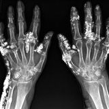
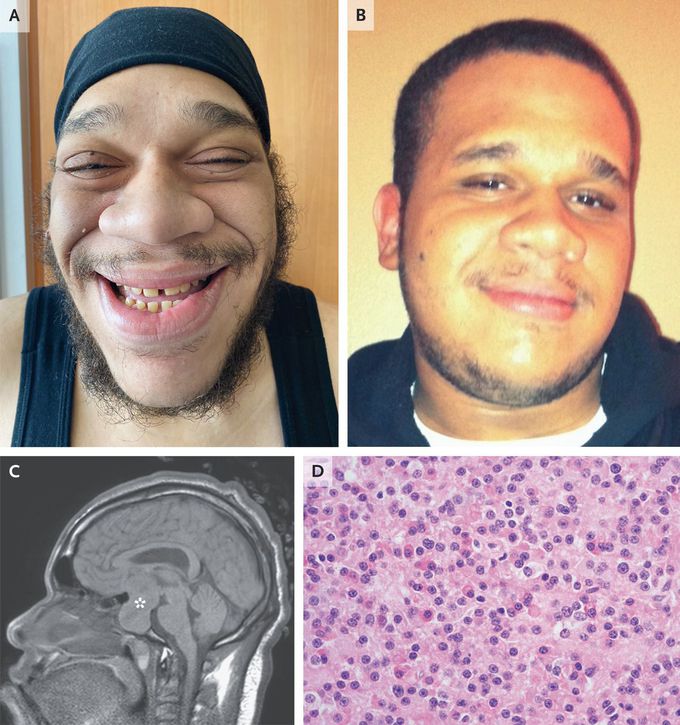
A 34-year-old man who had been admitted to the hospital with diabetic ketoacidosis was noted to have coarse facial features. He had no known medical history but had noted an increase in shoe size by three full sizes over the past 10 years and blurry vision for the past 3 years. Physical examination showed a widened nose, frontal bossing, prominent lower jaw, and malocclusion of the upper incisors (Panel A), findings not seen in a photograph taken 15 years earlier (Panel B). Visual-field testing showed bitemporal hemianopsia. Laboratory studies showed a glycated hemoglobin level of 12.3% (reference range, 5 to 14), an insulin-like growth factor 1 (IGF-1) level of 478 ng per milliliter (reference range, 53 to 331), and a growth hormone level of more than 65 ng per milliliter (reference value, ≤7.1). A diagnosis of acromegaly was made. Magnetic resonance imaging of the head revealed a pituitary macroadenoma measuring 6.0 cm by 3.4 cm by 2.8 cm (Panel C, asterisk) and macroglossia. Transsphenoidal partial tumor resection was performed. Histopathological testing showed a somatotroph adenoma (Panel D, hematoxylin and eosin staining) that also stained positive for growth hormone. The patient was discharged with instructions to receive monthly octreotide to treat residual tumor and with medications for diabetes and hypertension. At 1 month of follow-up, the patient’s IGF-1 level had reduced, but the mass gradually reexpanded, which led to a second resection 8 months after the initial surgery. Joshua Weinreb, M.D., Ph.D. Albert Einstein College of Medicine, Bronx, NY source: nejm.org

Living with HIV was one of the hardest experiences of my life. The fatigue, the emotional toll, and the uncertainty about the future weighed on me every single day. I had tried many treatments and medications, but nothing seemed to restore my health or energy the way I hoped.Out of both hope and desperation, I came across NaturePath Herbal Clinic. At first, I was skeptical but something about their natural approach and the powerful stories I read gave me the courage to try one more time.I began their herbal treatment program, and within a few weeks, I noticed small but meaningful changes more energy, better sleep, and a stronger immune system. Over the months, those improvements only grew. Today, I can truly say my life has changed. I feel healthier, more balanced, and finally in control of my well-being again.This isn’t just a testimony it’s a heartfelt recommendation to anyone living with HIV or any chronic condition. Don’t give up hope. I’m so grateful I gave NaturePath Herbal Clinic a chance. Visit their website to learn more: www.naturepathherbalclinic.com Email: info@naturepathherbalclinic.com
