


Anterior triangle of neck
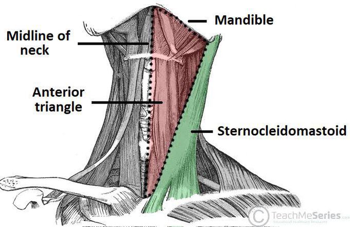
The anterior triangles refer to bilateral anatomic subdivisions of the neck comprising the anterior surface of the neck, deep to the superficial cervical fascia and platysma muscle. Laterally, the anterior triangle is bounded by the anterior border of the sternocleidomastoid muscle. Its superior border is the inferior border of the mandible. Medially, the boundary is the midline of the neck. The anterior triangle can further subdivide into four sub-triangles. The submandibular and submental triangles are the superior divisions, while the muscular and carotid triangles compose the inferior divisions of this anterior neck compartment. The submandibular triangle is delineated by the inferior border of the mandible and the anterior and posterior bellies of the digastric muscle. The submental triangle is demarcated by the hyoid bone, the anterior belly of the digastric muscle, and the midline of the neck.[1] The muscular triangle is outlined by the anterior aspect of the sternocleidomastoid and superior belly of the omohyoid, the midline of the neck, and the hyoid bone. The outline of the carotid triangle is the anterior aspect of the sternocleidomastoid, the superior belly of the omohyoid, and the posterior belly of the digastric and stylohyoid muscles.[2] The investing layer of the deep cervical fascia encases all of the infrahyoid muscles and forms the superficial boundary of the entire anterior triangle. The deep boundary of the carotid and muscular triangles is composed of the pretracheal layer of the deep cervical fascia, while the mylohyoid muscle forms the deep boundary of the submandibular and submental triangles.[3] The submandibular gland, a major salivary gland, emerges from the lateral aspect of the mylohyoid muscle and occupies most of the submandibular triangle.
