Andy Wellsover 5 years ago
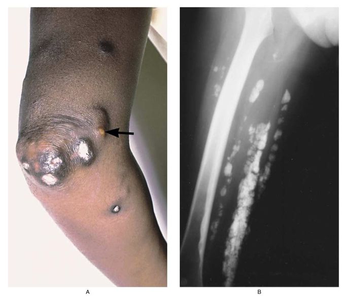
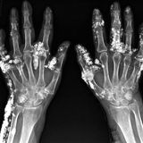
Calcifications in Dermatomyositis
The elbow (Panel A) of a 33-year-old woman who had had dermatomyositis since childhood shows subcutaneous calcifications extruding through the skin. A focus of purulent inflammation within a calcium deposit is evident (arrow). An x-ray film of the patient's thigh (Panel B) shows calcifications beneath the skin and within the muscle tissue. Some lesions on the skin in this area were palpable, and others were not visible. Deposition of calcium continued in this patient despite therapy with prednisone, azathioprine, and diphosphonates. Marinos C. Dalakas, M.D. National Institute of Neurological Disorders and Stroke, Bethesda, MD 20892 source: nejm.org
Other commentsSign in to post comments. You don't have an account? Sign up now!