


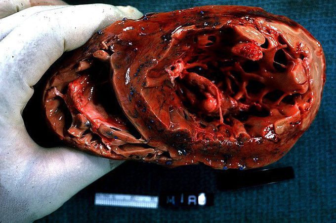
Papillary muscle infarct with rupture!!
This is a gross horizontal section of the left ventricle looking toward the base of heart. A great example of lateral wall transmural infarct and ruptured papillary muscle. Broadly speaking, the lateral wall of the LV is supplied by branches of the left anterior descending (LAD) and left circumflex (LCx) arteries. Infarction of the lateral wall usually occurs as part of a larger territory infarction, e.g. anterolateral myocardial infarction, and is caused by thrombosis in the artery. Complications of acute myocardial infarction (MI) include ischemic, mechanical, arrhythmic, embolic, and inflammatory disturbances. Nevertheless, circulatory failure from severe left ventricular dysfunction or one of the mechanical complications of MI account for most fatalities. There are three catastrophic mechanical complications of acute MI. These include left ventricular free wall rupture, rupture of the interventricular septum, and papillary muscle rupture. All involve loss of structural integrity of the infarcted tissue and are associated with extraordinarily high mortality rates if not promptly recognized and treated. Papillary muscle rupture frequently presents with symptoms ranging from acutely decompensated heart failure to cardiogenic shock. The most frequent scenario involves infarction upstream from the posterior descending artery (i.e. the right coronary artery in right dominant systems or the left circumflex artery in left dominant systems). Valvular competence during ventricular systole is maintained by the actions of two papillary muscles (anterolateral and posteromedial). The anterolateral muscle typically has a dual blood supply while the posteromedial muscle is supplied from only the PDA. Therefore the posteromedial muscle is more susceptible to infarction and rupture. Rupture leads to acute mitral valve regurgitation, pulmonary edema, and cardiogenic shock. Treatment often necessitates emergent surgical intervention, with mitral valve repair (if muscle necrosis is limited) or valve replacement. Photo by Peter Anderson.
