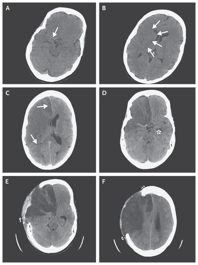


Evolving Infarction in the Anterior Circulation
A 47-year-old woman with a history of migraine was admitted to the hospital with acute-onset headache on the left side of her head and mild weakness of her left arm. Computed tomography (CT) of the brain without the use of contrast material was performed 5 hours after symptom onset and showed linear areas of hyperdensity in the first segment of the right middle cerebral artery (Panel A, arrow) and the first segment of the anterior cerebral artery. There were early signs of cerebral edema, including subtle low attenuation with loss of gray–white differentiation and effacement of sulci in both the right middle-cerebral-artery and anterior-cerebral-artery territories (Panel B, arrows). Four hours later, the patient became drowsy, and complete paralysis of the left side developed. A CT scan of the brain 9 hours after symptom onset showed large, nonhemorrhagic infarcts in the middle-cerebral-artery and anterior-cerebral-artery territories, with increased edema, mild right-to-left midline shift (Panel C, arrows), and some trapping of the left lateral ventricle (Panel D, asterisk). A total of 21 hours after symptom onset, the patient underwent decompressive craniectomy of the frontoparietal region (Panels E and F). She had a gradual recovery but died 6 weeks later from pneumonia.

