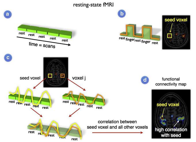


Resting-State functional Magnetic Resonance Imaging (RS-fMRI) studies are focused on measuring the correlation between spontaneous activation patterns of brain regions. Within a resting-state experiment, subjects are placed into the scanner and asked to close their eyes and to think of nothing in particular, without falling asleep. Similar to conventional task-related fMRI, the BOLD fMRI signal is measured throughout the experiment (panel a). Conventional task-dependent fMRI can be used to select a seed region of interest (panel b). To examine the level of functional connectivity between the selected seed voxel i and a second brain region j (for example a region in the contralateral motor cortex), the resting-state time-series of the seed voxel is correlated with the resting-state time-series of region j (panel c). A high correlation between the time-series of voxel i and voxel j is reflecting a high level of functional connectivity between these regions. Furthermore, to map out all functional connections of the selected seed region, the time-series of the seed voxel i can be correlated with the time-series of all other voxels in the brain, resulting in a functional connectivity map that reflects the regions that show a high level of functional connectivity with the selected seed region (panel d) (M.P. van den Heuvel, H.E. Hulshoff Pol, 2010)

