


Vertebral-Body Erosion in Thoracic Aortic Aneurysm
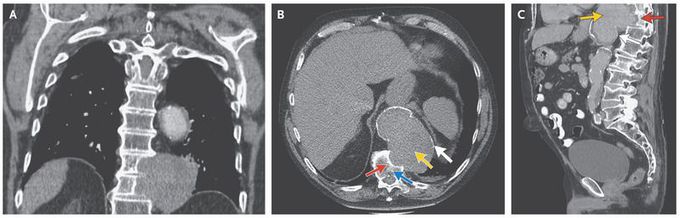
A 74-year-old man presented with acute back pain but without neurologic symptoms. The blood pressure was normal. He had a history of hypertension, open repair of an aortic infrarenal aneurysm, and end-stage renal failure that required hemodialysis. Computed tomography revealed a thoracic aneurysm that measured 8.1 cm by 11.7 cm in the greatest dimensions on the axial view and well-corticated erosions of thoracic vertebrae 10 and 11 (Panel A shows the coronal view, Panel B the axial view, and Panel C the sagittal view; yellow arrows show the aneurysm, red arrows the erosion, and the blue arrow thoracic vertebra 11). Thoracic vertebral erosion, which is more often seen after aortic graft surgery, is a rare complication of thoracic aortic aneurysm. The suggested mechanism is repetitive mechanical pressure causing relative ischemia in the bone, which leads to lysis and bone destruction. Differential considerations for a retroperitoneal mass eroding vertebrae include tumor and infection. The preservation of disk spaces seen in this patient makes infection unlikely, and the presence of a thin calcific rim surrounding the mass (Panels B and C, white arrows) is most consistent with a large aortic aneurysm. Because of the patient’s poor general health, no surgical repair was performed. With conservative treatment, the patient lived another 3 years. He died after a short episode of recurrent back pain for which palliative treatment was given.
