


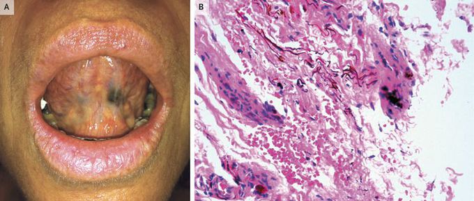
Oral Amalgam Tattoo Mimicking Melanoma
A 49-year-old woman without a personal or family history of melanoma presented to the dermatology clinic with an asymptomatic pigmented lesion on the ventral surface of her tongue. The lesion, which had been present for 4 months, was suggestive of oral melanoma. The patient reported no trauma and no alcohol or cigarette use. However, she did report that she had undergone a dental restoration procedure 3 months before the appearance of the lesion. Physical examination revealed an asymmetric, brownish-gray pigmented macule, measuring 1.0 cm by 1.2 cm in the greatest dimensions, with a central area of regression (Panel A; the white material in the patient’s mouth was chewing gum). No other cutaneous or mucosal abnormalities were found. The lesion was completely excised. Histopathological examination revealed dermis with fibrosis and an interstitial deposit of thin, elongated black pigment, with no inflammatory-cell infiltrate (Panel B; hematoxylin and eosin staining). No melanocytic hyperplasia or evidence of cancer was found. A diagnosis of amalgam tattoo was made. After 1 year of follow-up, there was no evidence of recurrence.
