


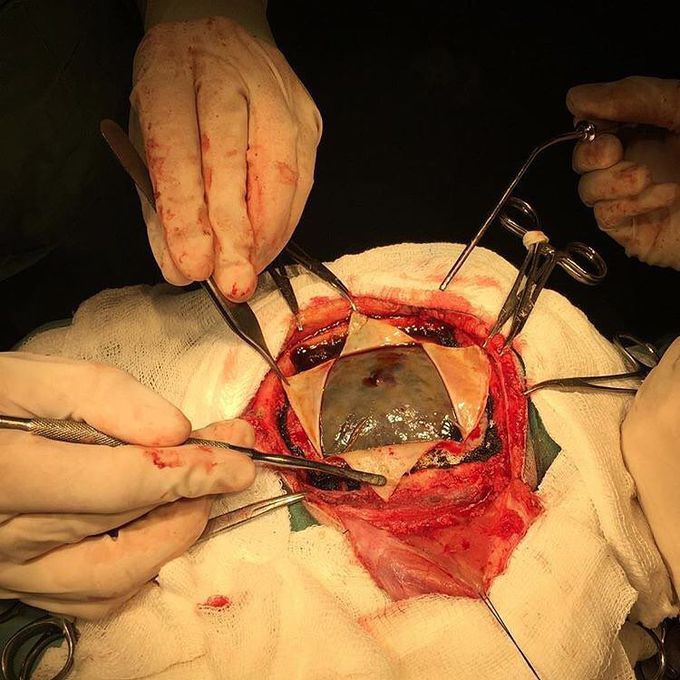
Look at that hematoma bulging out!
A subdural hematoma is among the deadliest of all head injuries. It results in blood gathering between the inner layer of the dura mater and the arachnoid mater, usually from tears in bridging veins which cross the subdural space. The bleeding fills the brain area very rapidly, compressing brain tissue. All the built up pressure due to the intra-cranial bleeding pushes the hematoma out once craniotomy is done - hence why it's bulging. It occurs not only in patients with severe head injury but also in patients with less severe head injuries, particularly those who are elderly or receiving anticoagulants. In a chronic subdural hematoma, small veins on the outer surface of the brain may tear, causing a gradual and slow bleeding in the subdural space, which can hide symptoms for several days or weeks.Small subdural hematomas can be managed by careful monitoring until the body heals itself. Other small subdural hematomas can be managed by inserting a temporary small catheter through a hole drilled through the skull and sucking out the hematoma. The big ones require surgery.
