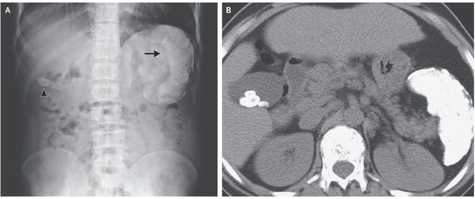


Calcified Spleen and Gallstones
A 35-year-old man presented to the gastroenterology clinic for treatment of chronic hepatitis C virus (HCV) infection. He had a type of sickle cell disease, hemoglobin Sβ thalassemia, with recurrent, episodic abdominal pain that had been present since childhood. A complete blood count showed microcytic anemia. The total bilirubin level was 8.5 mg per deciliter (145 μmol per liter; reference range, 0.3 to 1.2 mg per deciliter [5.1 to 20.5 μmol per liter]) and the indirect bilirubin level was 6.2 mg per deciliter (106 μmol per liter; reference range, 0.1 to 1.0 mg per deciliter [1.7 to 17.1 μmol per liter]). A peripheral-blood smear showed microcytic and hypochromic red cells, target cells, nucleated red cells, and sickle cells. An abdominal radiograph, which was obtained during a previous presentation for abdominal pain, showed a radiopaque gallstone (Panel A, arrowhead) and a calcified spleen (Panel A, arrow). A computed tomographic scan of the abdomen, obtained without the administration of contrast material, showed multiple gallstones and a calcified splenic pulp and capsule (Panel B). Pigment gallstones may occur as a result of hemolysis. Bilirubin stones, which are normally radiolucent, can be radiopaque when bilirubin binds with calcium. This patient completed treatment for chronic HCV infection, and he was vaccinated against encapsulated organisms. He has continued to do well on follow-up visits. Ankur Gupta, D.M. Priyanka Jain, M.D. Max Super Specialty Hospital, Dehradun, India source: nejm.org
I was diagnosed as a Hepatitis B carrier in 2015, with early signs of liver fibrosis. At first, antiviral medications helped control the virus but over time, resistance developed, and the effectiveness faded. I began to lose hope. In 2021, I discovered NaturePath Herbal Clinic despite my skepticism, I decided to give their herbal treatment a try.To my surprise, after just six months, my blood tests came back negative for the virus.It was nothing short of life-changing.I never expected such incredible results from a natural treatment. But it not only cleared the virus it restored my hope, my health, and my peace of mind.If you or someone you know is battling Hepatitis B, I truly encourage you to explore the natural healing path offered by NaturePath Herbal Clinic. It gave me a second chance and it might do the same for you.www.naturepathherbalclinic.com info@naturepathherbalclinic.com


