Clinical case
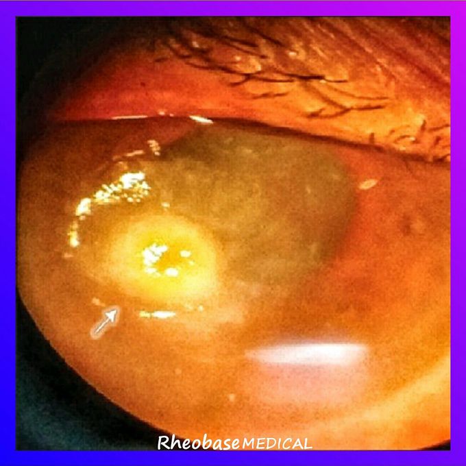
Corneal ulcers can be confusing, so I thought that I'll post another case to help you with getting a better understanding on how to differentiate Corneal ulcers. So lets get right into the case report. 👇👇 Q. A 65-year-old female 👵 on immunosuppressants 💊 for organ transplant presented with mild pain, redness, watering, and photophobia in the right eye (oculus dexter; OD) for the past 8 days. There was a history of trauma with cashew nut while working at home 🏡, following which the ocular symptoms developed. . Ocular 👀 examination revealed normal eyelids and diffusely congested conjunctiva. Slit lamp biomicroscopy of the cornea revealed an ulcer (1.4 mm × 1.2 mm) in the paracentral zone at 8 clock hours. The ulcer had a brownish pigmented plaque on the surface, with surrounding stromal infiltrate (4.8 mm × 2.5 mm) involving anterior two-third stroma with feathery extensions. Descemet's membrane folds and a faint immune ring were also seen. Anterior chamber did not have any hypopyon. Lens was cataractous. . What is your diagnosis? 💭