
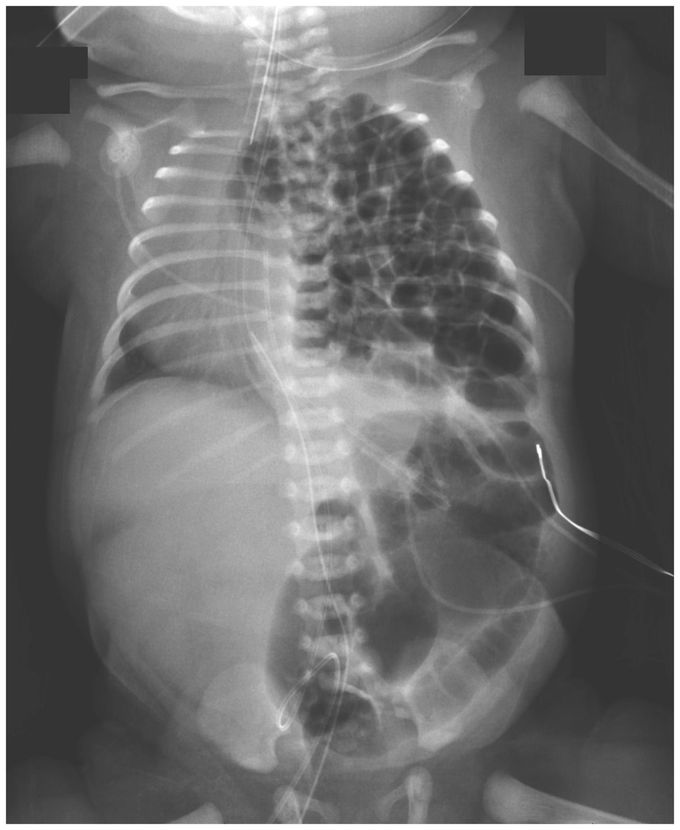
Congenital Diaphragmatic Hernia
A female infant was delivered by planned cesarean section at 36 weeks of gestation because of a prenatal diagnosis of congenital diaphragmatic hernia. The neonate was intubated immediately after delivery. A nasogastric tube was inserted and suction begun to decompress the bowels to allow for better lung expansion. Chest radiography revealed multiple loops of bowel occupying the left hemithorax, which shifted the cardiothymic structures to the right. Congenital diaphragmatic hernia occurs when the diaphragm muscle fails to close during fetal development; the defect can occur on the right side, left side, or, on rare occasions, both sides. Defects on the right side, which manifest with the liver in the chest, are treated with diaphragmatic patch repair and are associated with higher rates of high-frequency ventilation, extracorporeal membrane oxygenation, and death than are defects on the left side. In our patient, the posterolateral diaphragmatic defect, also called a Bochdalek hernia, measured 3 cm by 4 cm, and because of the more favorable location on the left side, the defect was fixed surgically by primary repair on the third day of life. After the surgery, the patient received high-frequency ventilation but not extracorporeal membrane oxygenation. She was discharged home from the neonatal intensive care unit after 1 month and did well. However, the defect recurred 6 months later, and a second operation was performed. Jared Klein, M.D. Megan Sirota, M.D. Children’s Hospital of Richmond at Virginia Commonwealth University, Richmond, VA source:nejm.org