


Zunaira saleh11 months ago
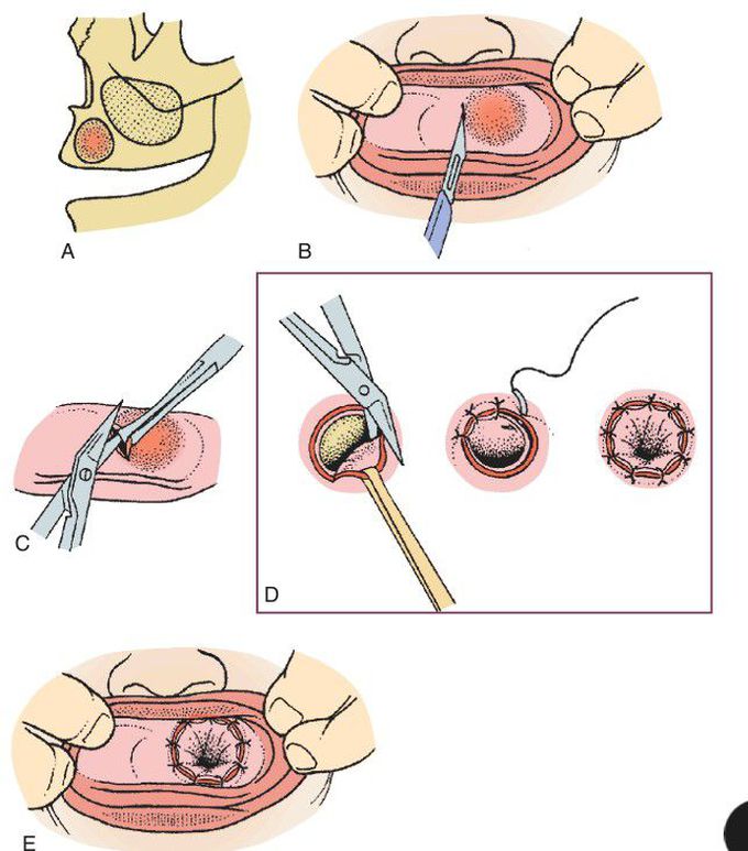
Marsupialization technique.
(A) Cyst within the maxilla. Palpation of the mucosa often reveals a compressible firmness, indicating that the bone has been eroded. The undersurface of the oral mucosa and the undersurface of the cyst (fibrous) capsule will therefore be fused together. (B) Incision through the oral mucosa and cystic wall into the center of the cyst. (C) Scissors are used to complete excision of the window of mucosa and cystic wall. This specimen is submitted for pathologic examination. (D) Oral mucosa and mucosa of the cystic wall sutured together around the periphery of the opening. This effectively “decompresses” the cyst, and it will now shrink as new bone fills in the cystic cavity.
Other commentsSign in to post comments. You don't have an account? Sign up now!
