


Zunaira saleh12 months ago
Cbct case study
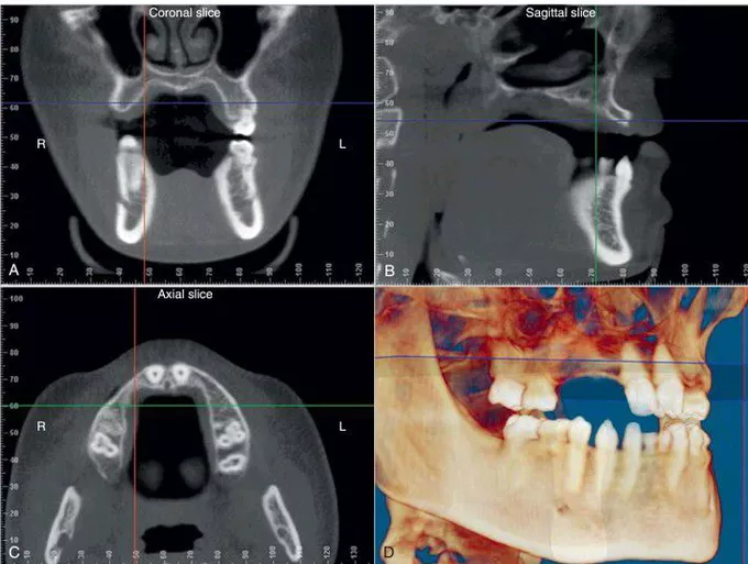
Cone-beam computed tomography scan allowing visualization of multiple structures in three dimensions. (A) The coronal slice through the posterior edentulous area demonstrating anatomy of the maxillary sinus and alveolar ridge bone. (B) A cross-sectional view of the edentulous anterior maxillary ridge. (C) An axial view showing deficiency of the anterior maxillary ridge. (D) Three-dimensional reconstruction.
Other commentsSign in to post comments. You don't have an account? Sign up now!
