
Zunaira salehabout 1 year ago

Tooth
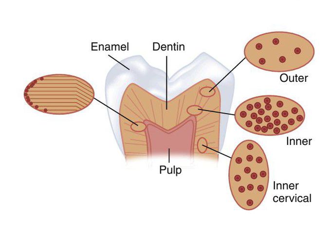
Schematic diagram of a tooth cut longitudinally to expose the enamel, dentin, and the pulp chamber. On the right side are illustrations of dentin tubules as viewed from the top, which shows the variation in the tubule number with location. At the left is an illustration of the change in direction of the primary dentin tubules as secondary dentin is formed
Other commentsSign in to post comments. You don't have an account? Sign up now!

