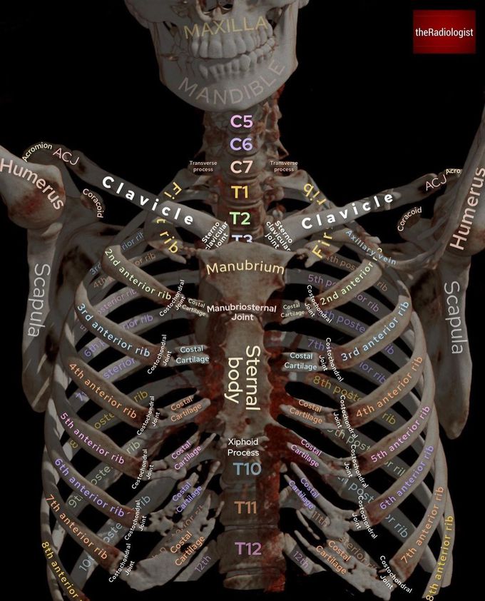


3D CT Thorax I
Have a look at this 3D reconstruction of the bones from a CT thorax and swipe left to compare this to how we see these structures on a Chest X-Ray Let’s have a think about the sternum: it is made up of three parts 1️⃣Manubrium: contains two clavicles notches and articulates with the medial end of each clavicle at the sternoclavicular joint 2️⃣Body: contains facets on its lateral borders to articulate with the costal cartilage of the 3rd-7th rib. Articulates with the second rib at the sternal angle 3️⃣Xiphoid process: has a variable shape Sternal fractures can result from blunt anterior chest wall trauma and deceleration injuries. These are not well seen on frontal X-Ray but we are picking more and more up on trauma CT scans. When reviewing on a sagittal view, the sternum is always a key review area for me as fractures can be difficult to appreciate on axial views.

