

Zunaira saleh5 months ago
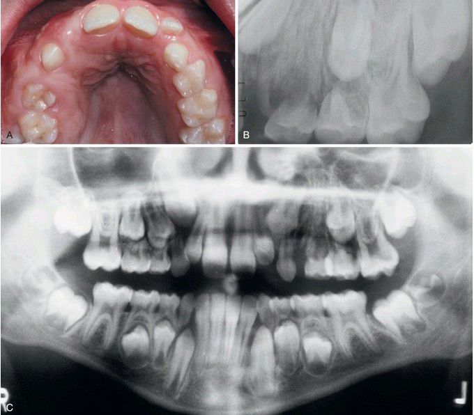
Segmental Odontomaxillary Dysplasia.
A, Unilateral enlargement of the maxilla and overlying gingival soft tissues. B, Periapical radiograph showing coarse trabecular pattern with absence of the first premolar. C, Panoramic radiograph showing irregular bone pattern of the left maxilla expanding into the maxillary sinus.
Other commentsSign in to post comments. You don't have an account? Sign up now!
Related posts
Florid Cemento-Osseous Dysplasia.Ectodermal DysplasiaEctodermal DysplasiaEctodermal DysplasiaHereditary Mucoepithelial Dysplasia.Hereditary Mucoepithelial DysplasiaUterine cervical dysplasiaLow-grade squamous intraepithelial lesion (L-SIL)High-grade squamous intraepithelial lesion (H-SIL)Renal cystic dysplasia.