


Case_ Study
What is the diagnosis? Chronic aortic dissection Cirrhosis Hypoplastic right heart syndrome Superior vena caval obstruction Transposition of the great vessels

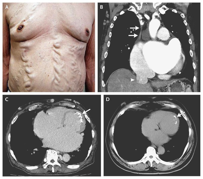
physical examination revealed varices of the chest and abdominal wall (image a)which may be caused by collateral circulation associated with obstruction of the superior vena cava (SVC). Chest (ct) showed SVC obstruction and dilatation of the inferior vena cava (image b) Endocardial calcification is obviously seen on image c so it is a svc obstruction...

The observed varices of the abdominal region is called “Caput Medusae”.

There are varices of the chest and abdominal wall caused by collateral circulation associated with obstruction of the superior vena cava. Paraumbilical and abdominal-wall varices can complicate cirrhosis but do not typically extend to the chest wall.

Phineas Gage: A Case Study in Brain Injury and Personality Change
A 70-year-old man presents with difficulty walking, particularly when turning, and a sensation of his feet being "stuck" to the floor. His gait is characterized by hesitation and freezing when initiating steps. Which of the following is most likely to be observed in this patient? A. Spasticity B. Foot drop C.Freezing of gait D. Romberg signBest Probiotics For Womens 2025: Are They More Effective?Effects of sugar on teeth
