


Herpes Simplex Dendritic Keratitis
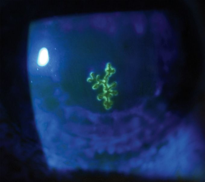
A 38-year-old man presented to the ophthalmology clinic with a 2-week history of tearing, photophobia, and reduced visual acuity in his left eye. Two years before presentation, he had undergone a deep anterior lamellar keratoplasty — also known as a partial-thickness corneal transplantation — on the left eye to treat advanced keratoconus. On ophthalmologic examination, the best corrected visual acuity of the left eye was limited to 20/80. A subsequent slit-lamp examination revealed mild conjunctival hyperemia and a branching epithelial ulcer with terminal bulbs involving the corneal graft. Fluorescein staining of the cornea that was viewed under illumination through a cobalt blue filter (see image) allowed further visualization of the dendritic ulcer. A diagnosis of herpes simplex dendritic keratitis was made. Keratitis is the most common form of ocular herpes simplex infection. The diagnosis is typically made on the basis of history and examination findings. Herpes simplex keratitis can complicate corneal transplants; therefore, patients who undergo corneal transplantation should be carefully monitored. Treatment with a course of topical 3% acyclovir ointment applied five times per day was initiated. At a 10-day follow-up visit, the patient’s symptoms had abated, the corneal graft had completely healed, and his best corrected visual acuity was restored to the baseline level of 20/25.
