


“Hairy Kidney” in Erdheim–Chester Disease
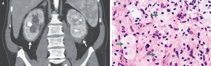
A 62-year-old man undergoing abdominal ultrasonography for the evaluation of gallstones was found to have a retroperitoneal mass. A physical examination and the results of routine laboratory studies, including tests of renal function, were normal. A computed tomographic (CT) urogram showed soft-tissue infiltration of the kidneys (Panel A, arrows), severe hydronephrosis of both kidneys, and moderate splenomegaly. Subsequent positron-emission tomography–CT showed no hypermetabolic activity. Stents were placed in both ureters, and a core-biopsy specimen of the perinephric soft tissue was obtained. Histopathological examination of the specimen revealed diffuse infiltration with pale-staining histiocytes (Panel B, arrows; hematoxylin and eosin staining) admixed with scattered lymphocytes and plasma cells. Immunostaining showed that the cells were strongly positive for CD68 and negative for S100 and CD1a. Molecular testing detected a BRAF V600E mutation. A diagnosis of Erdheim–Chester disease was made. Erdheim–Chester disease is a non–Langerhans-cell histiocytosis that is typically manifested by sclerotic lesions in the long bones — a feature that this patient uniquely did not have. This disease is also associated with numerous extraosseous findings, including infiltration of the retroperitoneal perinephric tissue, which results in a “hairy kidney” appearance on cross-sectional imaging, as was seen in this patient. Treatment with vemurafenib, a targeted inhibitor of the mutated BRAF V600E kinase, was started. At a 1-year follow-up visit, the patient had had no disease progression.
