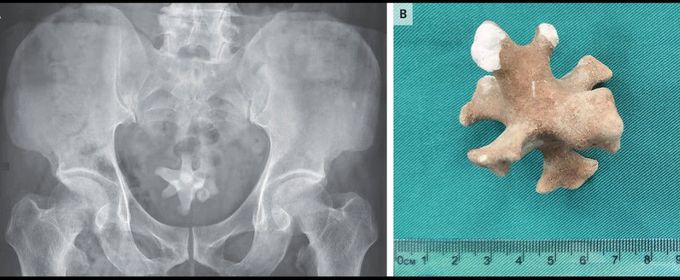


Jackstone Calculus in the Bladder
A 70-year-old man was referred to the urology clinic with a 3-month history of worsening symptoms in the lower urinary tract and new-onset hematuria. He had a history of recurrent urinary tract infections and benign prostatic hyperplasia, for which he had previously declined medications. However, 3 months earlier, he had started tamsulosin and finasteride to treat progressive dysuria, nocturia, frequency, and urgency. On physical examination, there was tenderness to palpation over the suprapubic region and an enlarged prostate. Urinalysis showed pyuria, hematuria, and bacteriuria. A urine culture grew extended-spectrum beta-lactamase–producing Escherichia coli, which was treated with ertapenem. Ultrasonography of the kidneys and bladder showed a small diverticulum in the posterior bladder wall and a large echogenic focus, indicating a possible bladder stone. A subsequent abdominal radiograph showed a large, irregularly shaped, radiopaque stone in the pelvis (Panel A). Transurethral enucleation of the prostate was performed, and a jackstone calculus measuring 5 cm in the largest dimension (Panel B) was removed from the bladder. Jackstone calculi — named after a toy jack — typically form in the bladder because of urine stasis and are composed of calcium oxalate dihydrate. At the 1-month follow-up, the patient’s symptoms had abated.

