


Wellens’ Syndrome
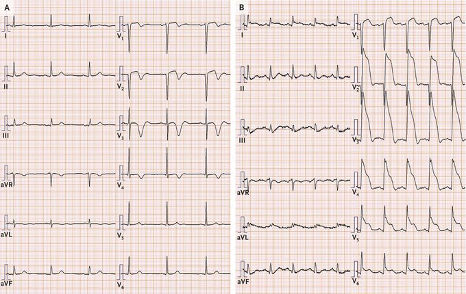
A 58-year-old man with coronary artery disease presented to the emergency department with a 1-day history of intermittent chest pain at rest. An electrocardiogram (ECG) that was obtained on arrival — at which time the patient reported no chest pain — showed biphasic T waves in leads V1 and V2 and inverted T waves in leads V3 and V4 (Panel A). The high-sensitivity troponin T level was 0.41 ng per milliliter (reference value, <0.1). Owing to concern about Wellens’ syndrome, the cardiology department was consulted, and catheterization was planned for later that day. Eighty minutes after presentation, the patient began having chest pain. A repeat ECG showed ST-segment elevations in leads V1 through V6 as well as in leads I and aVL (Panel B). Emergency coronary angiography identified a complete occlusion of the proximal left anterior descending artery, and a stent was placed Wellens’ syndrome refers to the biphasic or deep precordial T-wave inversions, particularly in leads V2 and V3, that are seen during a pain-free period that occurs after spontaneous reperfusion of an occluded left anterior descending artery. Rapid assessment and intervention are indicated, given the risk of coronary-artery reocclusion by an unstable plaque. The patient did well after the procedure and was discharged after a 2-week cardiac rehabilitation program.
