


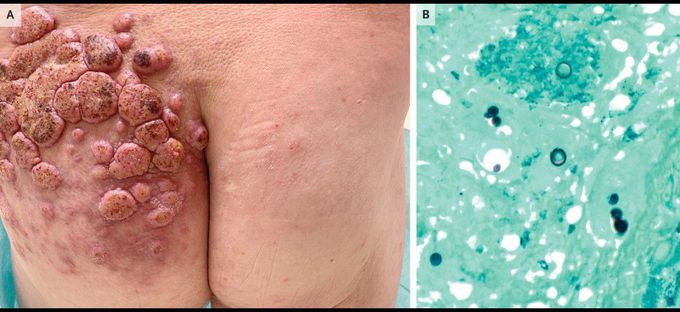
Primary Cutaneous Blastomycosis
A 53-year-old man presented to the dermatology clinic with a 4-month history of red, raised, itchy skin lesions on his left lower back and buttock. He had no known medical history and reported no constitutional or respiratory symptoms. He worked as a landscaper, often sustaining skin abrasions while trimming trees. The physical examination showed numerous verrucous nodules and plaques with overlying crusting and surrounding erythema on the left lower back and buttock (Panel A). Histopathological studies showed pseudoepitheliomatous hyperplasia with an intraepidermal and dermal infiltrate composed of neutrophils, lymphocytes, histiocytes, and eosinophils, as well as multinucleated giant cells with granuloma formation. Grocott–Gomori methenamine silver staining showed broad-based budding fungal organisms (Panel B), a finding consistent with Blastomyces dermatitidis. The result of a urine antigen test for blastomyces was also positive. Chest imaging showed no abnormalities. The patient received a diagnosis of primary cutaneous blastomycosis, which is caused by blastomyces species, dimorphic fungi found in the eastern half of the United States that typically grow in soil and detritus from wooded areas. Cutaneous manifestations usually indicate disseminated disease but rarely may occur by means of primary inoculation, as in this case. Itraconazole therapy was prescribed, and after 6 months the skin lesions had resolved.
https://www.facebook.com/PrimalTRTOfficial/Acute Abdomen Differential DiagnosisFlexiLeafhttps://www.facebook.com/StableGripSafetyBarOfficial/https://www.facebook.com/HumeHealthBodyPodOfficial/Pulsar Vexline Review 2026 - Legit Or Scam Trading Platform? Fact Check
