


Cerebral Cystic Echinococcosis
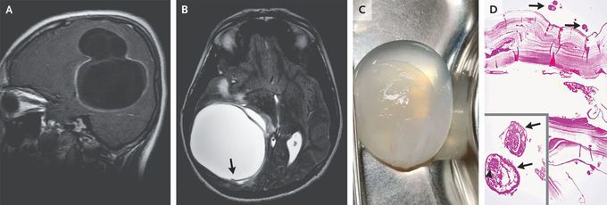
A previously healthy 14-year-old boy who lived on a farm presented with a 1-month history of episodic headaches associated with vomiting. Physical examination was notable for papilledema. Magnetic resonance imaging of the head showed a multiloculated cyst measuring 6.0 cm by 6.2 cm by 5.8 cm in the right temporoparietal region of the brain (Panel A, sagittal view, T1-weighted) with a hypointense rim and small projections in T2 phase (Panel B, axial view, arrow), findings suggestive of cystic echinococcosis. Computed tomography of the body showed no other sites of disease. A craniotomy was performed to excise the cyst. Saline irrigation was used to separate the cyst wall from the brain to avoid rupture (Panel C and video). Histopathological examination revealed an echinococcal laminated membrane lined by a germinal layer with daughter cysts (Panel D, arrows) and protoscolices (inset, arrows) with hooklets (arrowhead). A diagnosis of primary cerebral cystic echinococcosis from Echinococcus granulosus was made. Echinococcal cysts — also called hydatid cysts — most commonly form in the liver. The infection is transmitted to humans through contact with infected livestock or canine feces, as was likely to have happened in this case. Symptoms evolve if the cysts exert a mass effect. A 3-month course of albendazole was prescribed at discharge. At the 2-week follow-up, the patient’s symptoms had resolved. Sumit Thakar, M.B., B.S., M.Ch. Akhil Sunil, M.B., B.S., D.N.B. Sri Sathya Sai Institute of Higher Medical Sciences, Bangalore, India source: nejm.org
