


Sheeza Basharat6 months ago
Spinal Root Nerve
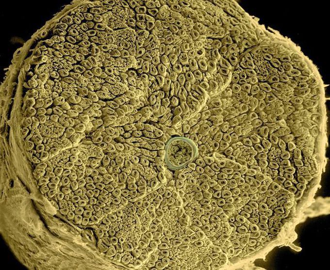
This is a scanning electron micrograph of a spinal root nerve showing the inner bundle of myelinated axons in cross-section. The entire nerve is about the diameter of a pencil lead. Photo Credit: Thomas Deerinck/ The National Center for Microscopy and Imaging Research, UCSD.
Source: https://www.instagram.com/p/Cno2jH6sTUu/?igshid=NDk5N2NlZjQ=Other commentsSign in to post comments. You don't have an account? Sign up now!
