


Cleft Palate
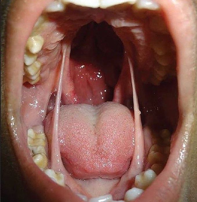
A 12-year-old boy was brought to the Out Patient Department for cleft palate. He was the first child of a healthy couple, born of nonconsanguineous marriage, and the antenatal period was uneventful. On examination he had a wide, complete bilateral cleft palate along with membranous bands connecting the margin of the palatal cleft with the floor of the mouth, lateral to the tongue bilaterally, which had been present since birth. These lateral oral synechiae were membranous, pink in color. They had broad attachments to the floor of the mouth measuring approximately 1.5 cm in width near the lingual side of first lower premolar tooth, and at the palatal margins (around hard palate-soft palate junction) the attachments narrowed to about 0.5 cm. The patient had no restriction of mouth opening. Examination of the ears, nose, extremities and genitalia did not reveal any anomaly. The developmental milestones were normal, and there was no evidence of mental retardation. Echocardiography and abdominal ultrasonography revealed normal visceral anatomy. The etiology of oral synechiae is unclear and several theories have been proposed. According to Longacre, oral synechia results from the persistence of the buccopharyngeal membrane. Kruger suggested that the mechanical effect of the tongue may contribute to the synechia between cleft palate margin and floor of the mouth. Mathis postulated that when adhesion of the palatal shelf occurs during developmental stages, adhering epithelial rudiments lead to oral synechia.
