

Achalasia
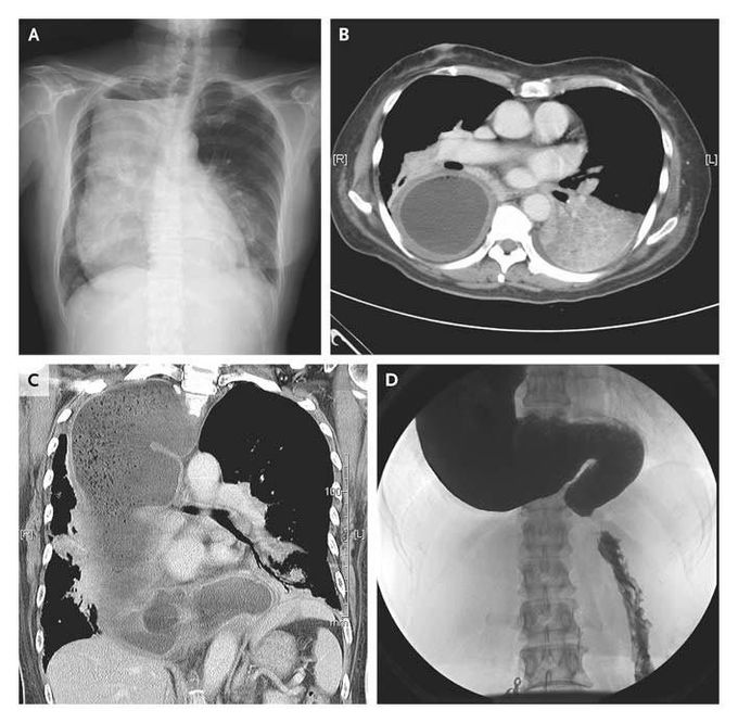
A 68-year-old woman presented with fever and dull pleuritic pain in the left chest wall. Chest radiography revealed a large mass with an air-fluid level in the right hemithorax (Panel A) and a suggestion of pneumonia in the left lower lobe. Computed tomography using contrast material revealed a severely dilated esophagus containing food, consolidation in the left lower lobe, and a compressed right lung (Panels B and C). The patient reported that she had had difficulty in swallowing food since her 20s and had adapted by eating a semiliquid diet for the past four decades. She reported often coughing when she lay in the left decubitus position. Achalasia was diagnosed by an upper gastrointestinal series (Panel D). She was successfully treated for aspiration pneumonia and was referred for further treatment of the achalasia. She declined surgical intervention as a possible means of improving the achalasia. Chi-Yen Liang, M.D. Ming-Shian Lin, M.D. Chia-Yi Christian Hospital, Chiayi City 600, Taiwan source: nejm.org

