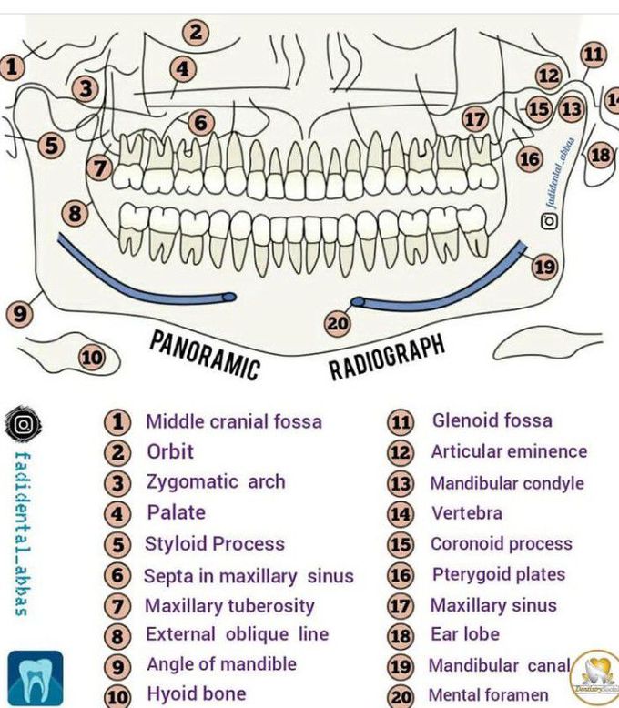


Panoramic X-ray
What is Panoramic X-ray? is a panoramic scanning dental X-ray of the upper and lower jaw. It shows a two-dimensional (2D) view of a half-circle from ear to ear. Panoramic radiography is a form of focal plane tomography; thus, images of multiple planes are taken to make up the composite panoramic image, where the maxilla and mandible are in the focal trough. ______________________________________________ What are some common uses of the procedure? A panoramic x-ray is a commonly performed examination by dentists and oral surgeons in everyday practice and is an important diagnostic tool. It covers a wider area than a conventional intraoral x-ray and, as a result, provides valuable information about the maxillary sinuses, tooth positioning and other bone abnormalities. This examination is also used to plan treatment for full and partial dentures, braces, extractions and implants. A panoramic x-ray can also reveal dental and medical problems such as: • advanced periodontal disease • cysts in the jaw bones • jaw tumors and oral cancer • impacted teeth including wisdom teeth • jaw disorders (also known as temporomandibular joint or TMJ disorders) • sinusitis ADVANTAGES: • Broad coverage of facial bone and teeth • Low patient radiation dose • Convenience of examination for the patient (films need not be placed inside the mouth) • Ability to be used in patients who cannot open the mouth or when the opening is restricted e.g.: due to trismus • Short time required for making the image • Patient's ready understandability of panoramic films, making them a useful visual aid in patient education and case presentation. By:https://www.instagram.com/p/CcdPyjsttFw/?utm_source=ig_web_copy_link

