


Closed Gastroschisis
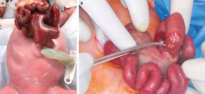
In a male neonate born at 37 weeks of gestation, a narrow stalk of the small bowel and proximal colon protruded through a 1.5-cm defect in the abdominal wall. A diagnosis of gastroschisis had been made antenatally on the basis of findings from prenatal ultrasonography. In the area where the bowel had traversed the defect, the jejunum was atretic and the colon was stenosed (Panel A). The term “gastroschisis” refers to the evisceration of the intestines through a defect in the abdominal wall; the condition is referred to as closed gastroschisis when the defect closes, causing incarceration of the eviscerated bowel. In this infant, laparotomy was performed on the first day of life. The cecum was found to be perforated (Panel B), and the proximal jejunum ended blindly within the abdomen. The colonic perforation was sutured, the bowels were reduced, and the abdomen was closed. An additional laparotomy was performed 19 days later to repair the jejunal atresia and colonic stenosis. After transitioning from parenteral to enteral nutrition at 27 days, the boy was discharged home, at 1 month of age. Tyler D. Robinson, M.D., M.P.H. Christine Whyte, M.D. Albany Medical Center, Albany, NY Source: nejm.org
