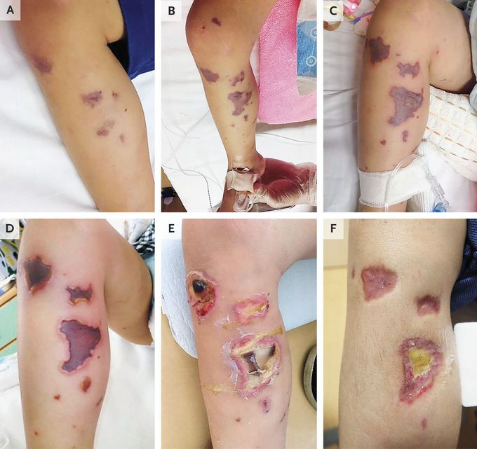
Evolution of Purpura Fulminans
A previously healthy 3-year-old boy presented to a children’s hospital with fever and vomiting. On physical examination, he had small purpura on his legs (Panel A) that spread to his face, abdomen, and limbs during the ensuing 3 hours. Treatment with broad-spectrum antibiotic agents was initiated immediately after blood specimens were obtained for culture. Over the next 6 hours, the purpura enlarged and spread further (Panel B), and signs of septic shock developed. Blood cultures grew Neisseria meningitidis, serotype B, confirming the diagnosis of acute infectious purpura fulminans caused by meningococcemia. Over the next 48 hours, the patient had clinical improvement and some smaller lesions receded, but lesions over the right wrist and right lower leg evolved into well-demarcated bullae with recessed centers (Panel C). Nine days after presentation, the lesions resembled ulcers (Panel D). The patient had difficulty moving his right leg smoothly because of pain and muscle weakness. With treatment, including wound care, the lesions diminished, with granulation tissue present at 3 weeks (Panel E). Four months after presentation, the lesions had epithelialized (Panel F), and the patient regained full range of motion in the leg. Tamae Kugai, M.D. Hidenori Nakagawa, M.D. National Center for Child Health and Development, Tokyo, Japan source: nejm.org