


Bilateral Nephrocalcinosis
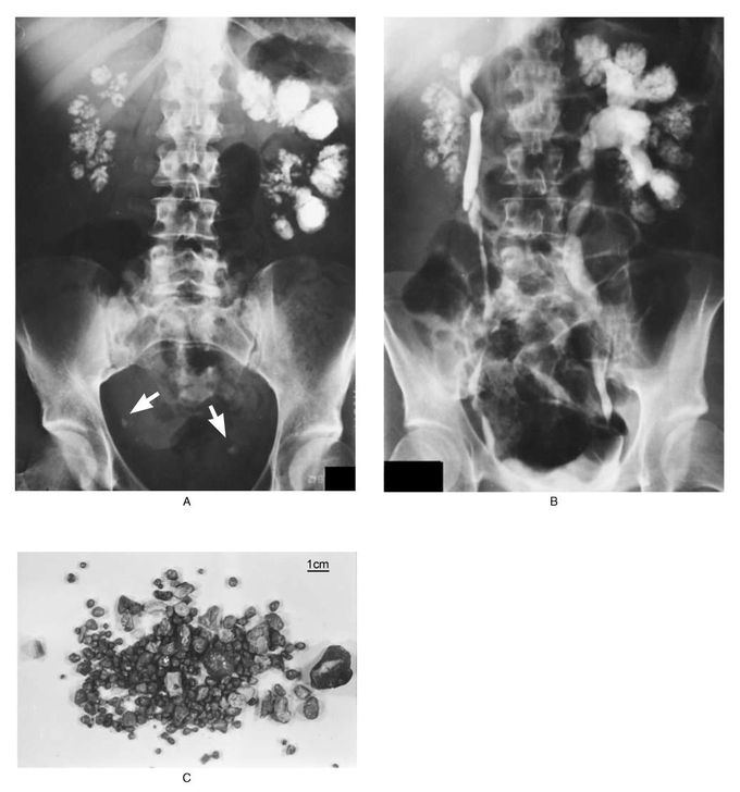
A 39-year-old woman presented with a seven-year history of renal calculi. A plain abdominal film obtained when she was 38 (Panel A) showed enlarged kidneys bilaterally with nephrocalcinosis in a papillary distribution and radiopaque calculi in both ureters (arrows). An intravenous urogram done at the same time (Panel B) showed left-sided hydronephrosis. A collection of stones passed by the patient over a two-month period is shown in Panel C. Analysis of the stones showed that the majority were composed of calcium oxalate, with some composed of magnesium ammonium phosphate. Urine cultures showed intermittent coliform infection, with no evidence of urea-splitting organisms. The patient's usual electrolyte values were as follows: serum sodium, 140 mmol per liter; potassium, 4.0 mmol per liter; chloride, 105 mmol per liter; and bicarbonate, 23 mmol per liter. Her 24-hour urinary calcium excretion (290 mg [7.2 mmol] per 24 hr) was at the upper limit of normal for our laboratory. Urinary pH was never below 6, and an ammonium chloride-loading test revealed an acidification defect consistent with the presence of type 1 renal tubular acidosis. Despite a high fluid intake (>3 liters per day) she continues to pass stones intermittently. Fortunately, her serum urea and creatinine concentrations have remained normal. Jonathan T.C. Kwan, M.D. Frank P. Marsh, M.A., F.R.C.P. Royal London Hospital, London E1 1BB, United Kingdom Source: nejm.org
