


Maryum Mehboobover 4 years ago
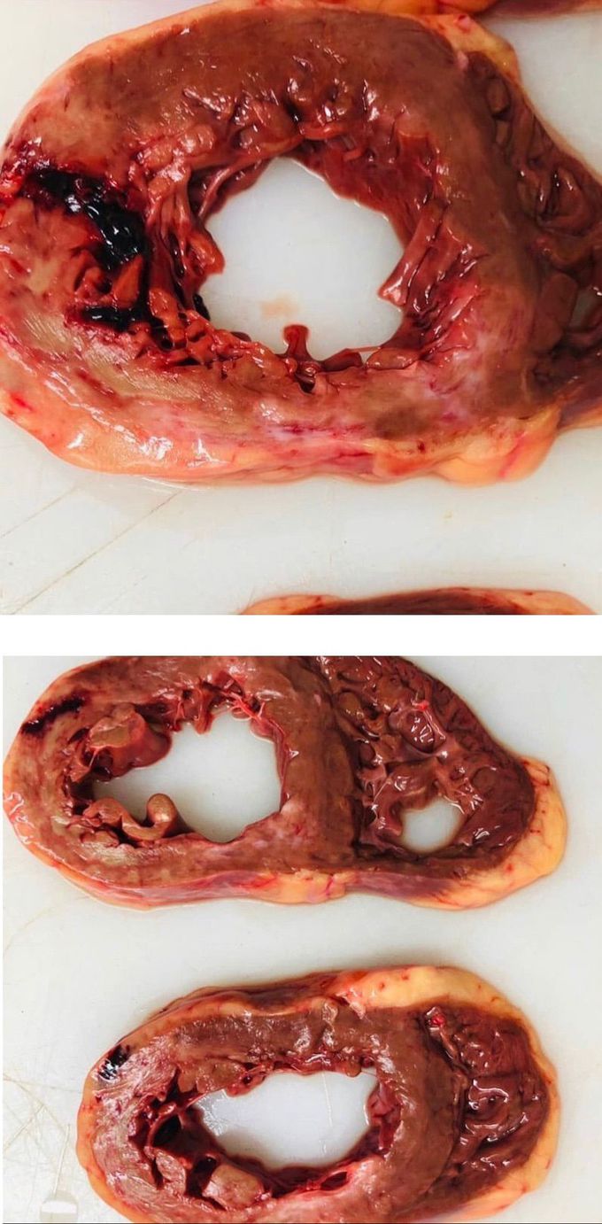
Ventricular rupture following MI
Ventricular rupture following MI This is the heart of a 79 year old male smoker showing rupture of the left ventricular wall following Myocardial infarction. Rupture of the free wall led to accumulation of the blood between the heart muscle and pericardium. This resulted in cardiac tamponade. Classical signs of cardiac tamponade are hypotension, distant heart sounds and jugular vein distension also called Beck’s triad. Picture credit: @themedicalmentors (via Instagram) https://www.instagram.com/p/CNh2p9MnaOY/?igshid=sa77a1qng4s1

Other commentsSign in to post comments. You don't have an account? Sign up now!
Related posts
Blue Baby Heart Defect: Tetralogy of FallotAngioplasty, Causes, Signs and Symptoms, Diagnosis and Treatment.HypertensionPremature Ventricular Complexes (PVCs)TachycardiaSupraventricular TachycardiaSupraventricular tachycardia- DiagnosisPatent Ductus Arteriosus- SymptomsAtrial Septal Defect- Diagnosis
