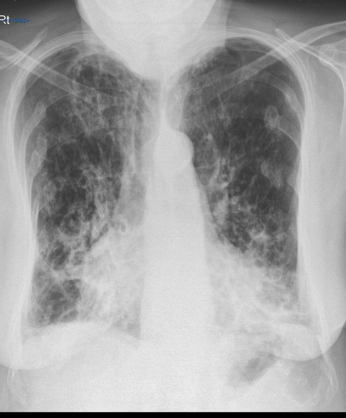


Radiological Findings in Bronchiectasis
Bronchiectasis refers to abnormal irreversible dilation of bronchial system usually secondary to infections and cystic fibrosis. Majority of cases are idiopathic. Patient with bronchiectasis presents with the history of chronic productive cough, recurrent chest infections, and hemoptysis. The investigation of choice for diagnosis of bronchiectasis is high resolution CT. However, characteristic Xray findings are often demonstrated. -Tram-Track Opacities These are usually seen in cylindrical bronchiectasis. -Air-Fluid Levels It is demonstrated in cystic bronchiectasis. -Increased Bronchovascular Markings -Ring Shadows of Bronchi -Ill-defined pulmonary vasculature It indicates peribronchovascular fibrosis. Reference: https://radiopaedia.org/articles/bronchiectasis
Thank you for sharing this marvelous film and the differential disease correlations.
Never seen it on an x-ray, so good to see what it might look like before CT
Living with Pulmonary Fibrosis (PF) was one of the hardest experiences of my life. The breathlessness, the fatigue, and the fear of the future weighed on me every single day. I had tried so many treatments and medications, but nothing seemed to stop the disease from progressing.Out of both hope and desperation, I came across NaturePath Herbal Clinic. At first, I was skeptical but something about their natural approach and the stories I read gave me the courage to try one more time.I began their herbal treatment program, and within a few weeks, I noticed small changes easier breathing, more energy, and a clearer mind. Over themonths, those improvements became more and more obvious. Today, I can truly say my life has changed. My lungs feel stronger, and my quality of life has returned in ways I didn’t think were possible.This isn’t just a testimony it’s a heartfelt recommendation to anyone struggling with PF or other chronic conditions. Don’t give up hope. I’m so grateful I gave NaturePath Herbal Clinic a chance. Visit their website to learn more: www.naturepathherbalclinic.com info@naturepathherbalclinic.com


