


Aelia Sarv Jahanalmost 2 years ago
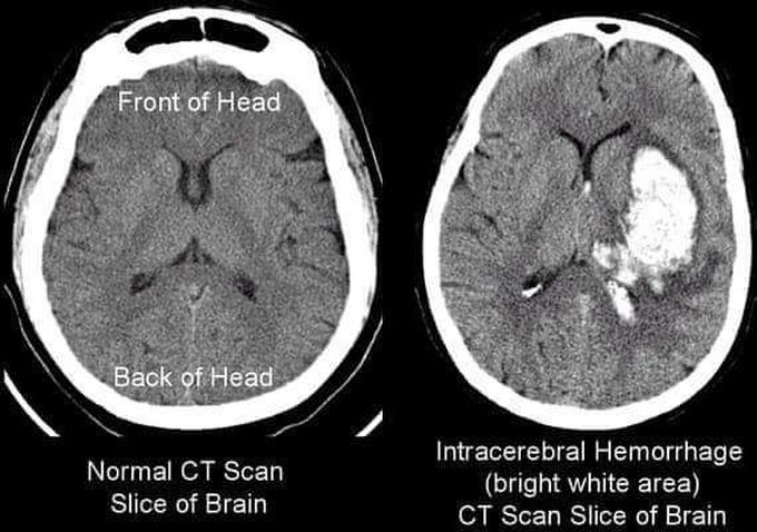
Your Consultant shows you a CT scan head during your rotation in the neurosurgery ward with findings as given above. What is your most likely diagnosis?
INTRACEREBRAL HEMORRHAGE: ✔Isolated hematomas within the brain parenchyma are most often associated with hypertensive hemorrhage or arteriovenous malformations (AVMs). Bleeding may occur in a contused area of brain. ✔Mass effect from developing hematomas may present as a delayed neurologic deficit. ✔Delayed traumatic intracerebral hemorrhage is most likely to occur within the first 24 hours. initial head CT scan should be reimaged 24 hours after the trauma to document stable pathology. ✔Indications for craniotomy include: ➖Any clot volume >50 cm3. ➖A clot volume >20 cm3 with referable neurologic deterioration (GCS 6–8). Associated midline shift >5 mm or basal cistern ➖compression. Source: Schawrt’z Principles of Surgery
Other commentsSign in to post comments. You don't have an account? Sign up now!
