


Zunaira saleh10 months ago
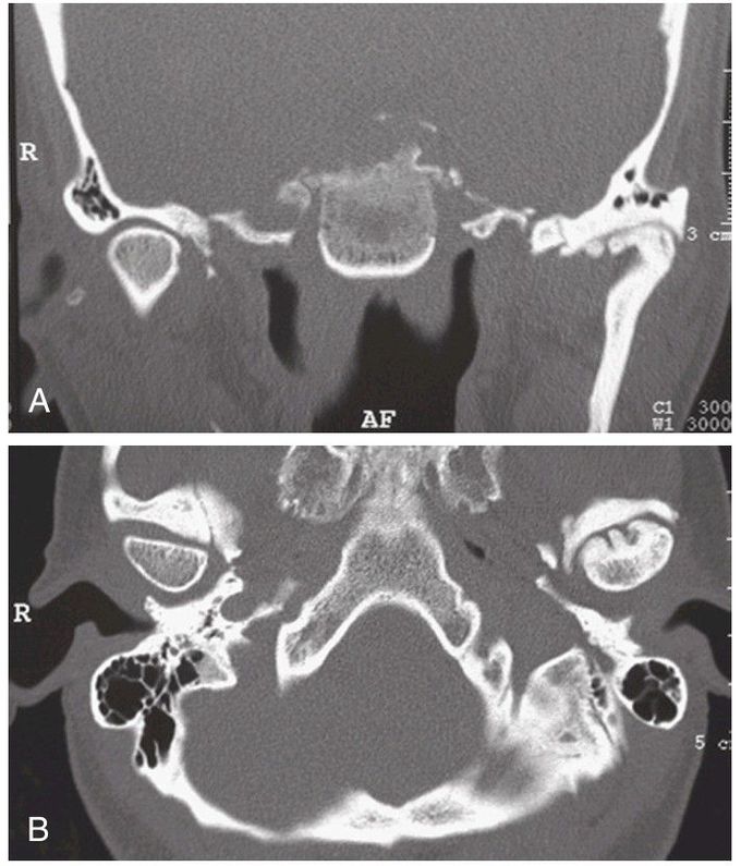
Computed tomography.
(A) Coronal image illustrates normal architecture of the right (R) condyle with alteration of the left condyle resulting from a history of trauma. (B) Axial view depicts the altered condylar anatomy referenced against the contralateral joint.
Other commentsSign in to post comments. You don't have an account? Sign up now!
