


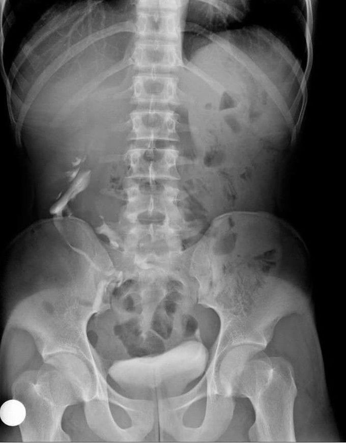
What's this intravenous urogram showing??
Epidemiology The estimated incidence is around 1 out of 1000 births 1. There is a recognised male predilection with a 2:1 male to female ratio. More than 90% of crossed renal ectopia results in fusion. Pathology It results as a consequence of abnormal renal ascent in embryogenesis with fusion of the kidneys within the pelvis. It is thought to occur in the first trimester, at around 4th-8thweek of fetal life (In a normal situation the kidney reaches its appropriate position at L2 level at the end of the 2nd month). Some evidence supports that an abnormally situated umbilical artery prevents normal cephalic migration. Another theory is that the ureteric bud crosses to the opposite side and induces nephron formation in the contralateral metanephric blastema. The result is a single renal mass with two collecting systems being located on one side of the abdomen. Normal ascent of the kidneys is required for formation of the extraperitoneal peri-renal fascial planes and therefore ectopia (or renal agenesis) results in failure of development of fascial layers in the flanks on the side not occupied by renal tissue. The lack of restraining fascia leads to possible malposition of bowel into the extra-peritoneal fat of the empty renal fossa and relaxation of mesenteric supports for bowel loops in this region. Subtypes type a: inferior crossed fusion type b: sigmoid kidney type c: lump kidney type d: disc kidney type e: L-shaped kidney type f: superiorly crossed fused Location Left-to-right ectopy is thought to be three times more common. Radiographic features Fluoroscopy Urography (IVU) The anomaly is readily detected on conventional urography. In 90% of crossed ectopy, there is at least partial fusion of the kidneys (the remainder demonstrate two discrete kidneys on the same side, crossed-unfused ectopy). An anterograde or retrograde ureterogram most often demonstrates normal bladder trigone without ureteral ectopy. Barium studies of the bowel Barium contrast studies of the bowel should be interpreted in light of bowel laxity in the region of the empty renal fossa (discussed above). In particular, distinction must be made from internal hernia. Ultrasound On ultrasound there may be a characteristic anterior or posterior "notch" between the two fused kidneys. CT The parenchymal band joining the two kidneys can be better visualized on CT scan. Also, anatomical relationship with adjacent structures and positions of the ureter can be better assessed. Complications In a crossed fused renal ectopic kidney, complications such as nephrolithiasis, infection, and hydronephrosis approaches ~50%. Treatment and prognosis Crossed fused ectopia usually doesn't require any primary treatment. However, understanding is essential before planning any surgical intervention in the renal region. The blood supply to cross-fused kidney is usually anomalous and angiography recommended before surgical intervention.
